|
Echinococcosis & Taeniasis |
 |
 |
 |
 |
|
| |
| Causative Agent |
|
Echinococcosis: |
|
|
|
Taeniasis: |
-
Parasitic disease of mammals caused by infection with
tapeworms of the genus Taenia.
-
Taenia ovis krabbei
is known to be present in BC.
-
Definitive and
intermediate hosts both are mammals.
-
Infection with Taenia
causes little or no harm to the definitive host; however, larval
stages within intermediate hosts can be
pathogenic.
|
| Images |
|
Click on images to enlarge. |
 |
 |
 |
|
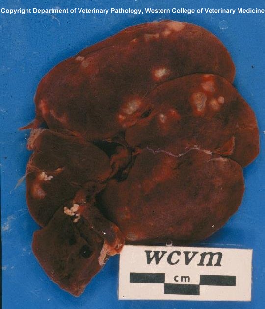
Cysts of
E. multilocularis
in the liver of a muskrat. |
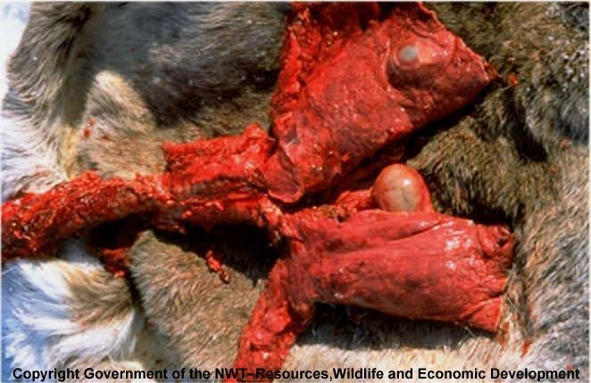
Caribou lungs with hydatid
cysts (E. granulosus). |
Generalized
Echinococcus life cycle. |
|
| Distribution |
|
Geographic: |
-
Echinococcus
granulosus is widely distributed
across Canada.
-
Echinococcus
multilocularis has a more limited
distribution, occurring in AB, MB, SK, and NWT. It recently
has been found in
BC
(PDF
link to journal article).
-
Taenia ovis krabbei
is found throughout Canada.
|
|
Seasonality: |
-
Carnivores act as
reservoirs all year long; herbivores
become infected by consuming contaminated vegetation when it
is available.
|
|
| Hosts, Transmission and Life
Cycle |
| Hosts: |
|
E.
granulosus |
-
Definitive (adult worm): wolves (Canis
lupus), coyotes (Canis
latrans), domestic dogs.
-
Intermediate (larval worm):
Cervids, particularly moose (Alces
alces), caribou (Rangifer
taradus), elk (Cervus
canadensis) and bighorn sheep (Ovis
canadensis) in western Canada.
-
Humans can act as
intermediate hosts but are considered to
be a dead-end host
since humans are not usually consumed by carnivores.
|
|
E. multilocularis |
-
Definitive (adult worm): Arctic and
red foxes (Vulpes),
coyotes, sometimes domestic dogs and cats.
-
Intermediate (larval worm): rodents,
such as voles (Cricetidae), mice (Muridae) and muskrats (Ondatra zibethicus).
-
Humans again can act as
intermediate hosts but are considered to
be a dead-end host
since humans are not usually consumed by carnivores.
|
|
Taenia ovis krabbei |
-
Definitive (adult worm): wolves,
coyotes, domestic dogs, cougars (Felis
concolor), bears (Ursidae).
-
Intermediate (larval worm):
Cervids such as moose, caribou, and
elk, and Rocky Mountain bighorn sheep.
|
|
Transmission and Life Cycle: |
|
Echinococcus |
-
See life cycle diagram above.
-
Life cycle is similar between E. granulosus and E.
multilocularis, with the main differences being host
species and larval growth characteristics.
-
Adult worms occupy the small intestine of infected carnivores and eggs
are voided in the feces, usually a month after initial
infection.
-
Eggs from the feces contaminate vegetation and are subsequently eaten by
intermediate, herbivorous hosts.
-
Larvae move to preferred
sites within the
intermediate host, usually lung or liver
(or less frequently, the muscle or eyes), where they form
often large and obvious fluid-filled hydatid
cysts containing many larvae.
-
Cysts are consumed by carnivores,
breaking open to release immature worms.
-
Larvae then mature into
adult worms after attaching to the wall of the small
intestine of the carnivore, subsequently releasing eggs within the feces.
|
|
Taenia
ovis krabbei |
-
Life cycle similar to species of Echinococcus.
-
Adult worms occupy the small intestine of carnivores as well as
omnivorous mammals, and are passed in feces.
-
In intermediate hosts (ungulates), larvae form
cysts, mainly in the skeletal
muscles and associated
connective tissues.
|
|
| Signs and Symptoms |
-
Adult worms have no detrimental effects on the carnivore host.
|
|
E.
granulosus |
-
Larval
cysts may cause problems in host
tissue because of the continual growth and expansion of the
cyst.
-
The structure of the wall of
the
cysts forms a tissue/host barrier
enabling tissues of the host to wall off the
cysts itself, preventing further
spread.
-
Subsequent compression of
tissues, such as the lung, may cause debilitation due to the animal’s reduced ability to breathe if a sufficient
number of
cysts are involved.
|
|
E. multilocularis |
-
More dangerous than E. granulosus as the larval
cysts grow rapidly and bud
externally, acting very much like an invasive cancer.
-
Unlike the
cysts of E. granulosus,
the structure of the wall of the
cysts of E. multilocularis
does not form a tissue/host barrier, allowing the
cyst to further invade tissues
via the
lymph or blood.
-
E. multilocularis severely
debilitates and often kills its rodent host.
|
|
Taenia ovis krabbei |
-
Larval forms have been associated with significant tissue damage and loss
of body condition in infected herbivores, but most infections are
noted by chance during butchering of hunter-killed animals.
|
| Meat Edible? |
-
Humans are not capable of harboring adult Echinococcus tapeworms
and so cannot become infected either by handling or eating hydatid
cysts - for aesthetic reasons,
cysts should be removed prior to
consumption.
-
Humans can, however, be
infected by consuming the infective eggs passed by the carnivore
hosts of Echinococcus. For this reason, those who handle live
carnivores, their feces, pelts or carcasses should wear gloves and
use good hygiene to avoid contamination by tapeworm eggs.
-
Taenia ovis krabbei
is not transmissible to humans during any part of its cycle;
cysts noted in meat are not
aesthetically pleasing but are killed during normal cooking
temperatures and by freezing.
-
Meat of animals infected by
these parasites should not be fed to dogs since they can be hosts
for the adult tapeworms. Also, infected viscera should be destroyed
by burning to prevent transmission to domestic dogs.
|
| Human Health Concerns and
Risk Reduction |
|
Echinococcus |
-
In humans, infection with E. granulosus is called hydatid disease
or cystic hydatid disease.
-
In humans, infection with E. multilocularis is called alveolar
hydatid disease.
-
Humans can become infected when feces of infected carnivores or carnivore
pelts that are contaminated with feces are handled, or from
environments contaminated with carnivore feces.
-
E. granulosus
infection in the lungs of humans may be associated
with fever and difficulty breathing.
-
E. granulosus
cysts may also develop in other
organs, including the brain, and cause severe problems because of
the pressure on normal tissue.
-
E. multilocularis
behaves like an invasive cancer and can cause liver damage resulting
in abdominal pain and jaundice; in areas where this parasite is
common, 70% of untreated cases become fatal within 5 years.
-
Human infections can be
treated with antiparasitic drugs or through surgical removal of
cysts.
-
Domestic dogs can serve as reservoirs for
Echinococcus infection within communities. They should not be
fed carcasses or allowed to scavenge from infected game mammals as
this perpetuates the cycle of infection.
-
Tapeworm infection in dogs and cats can be
treated with anthelmintics (drugs used against tapeworms).
-
Risk Reduction:
-
Always wear rubber gloves when handling carnivore pelts, droppings or
intestines.
-
Careful personal and food hygiene when in close proximity to dogs is
crucial in preventing human infection.
-
Eggs dry out easily and can die within 2 hours in direct sunlight;
survival time is increased in damp areas such as watering holes.
-
DO NOT FEED TISSUES
CONTAINING CYCSTS
TO DOGS.
|
|
Taenia ovis krabbei |
-
Taenia ovis krabbei is not transmissible to
humans.
|
| Samples for Diagnosis |
-
Tapeworm infection can be verified on the basis of finding eggs in the
fecal material of infected carnivores.
-
Cystic larval stages can be identified in
intermediate hosts on the basis of gross
appearance.
-
Human infection can be
verified by taking X-rays, CT scans, and through a variety of
immunological tests.
-
Portions of tissues
containing
cysts can be sent to appropriate
diagnostic laboratories.
|
| Further Reading |
-
Alberta Environment and Sustainable Resource Development –
Echinococcus (PDF file)
-
Alaska Department of Fish and Game –
Echinococcus
-
Alaska Department of Fish and Game – Muscle Tapeworm Cysts
-
Michigan Department of Natural Resources –
Echinococcus
-
Peregrine et al. 2012. Alveolar hydatid disease (Echinococcus
multilocularis) in the liver of a Canadian dog in British
Columbia, a newly endemic region. Canadian Veterinary Journal 53:
870-874. (PDF file)
-
US Centers for
Disease Control –
Echinococcus
-
World
Health Organization –
Echinococcus
-
Jones, A., and M. J. Pybus. 2001. Taeniasis and Echinococcosis. Pp.
150-192 in W. M. Samuel, M. J. Pybus, and A. A. Kocan (eds.),
Parasitic Diseases of Wild Mammals. 3rd Ed. Iowa State University
Press, Ames, IA.
-
Canadian Cooperative Wildlife Heath Centre. 1995. Echinococcosis. Pp.
37-38. Health risks to wildlife personnel: hazards from
disease-causing agents. Canadian Cooperative Wildlife Heath Centre,
Western College of Veterinary Medicine, University of Saskatchewan.
Saskatoon, SK
-
Elkin, B, and R. L. Zamke. 2001. Common wildlife diseases and parasites
in Alaska. Alaska Department of Fish and Game. Anchorage, AK.
|
|
|