| Besnoitiosis |
 |
 |
|
| |
| Causative Agent |
-
Besnoitiosis is caused by the unicellular
protozoan parasite Besnoitia
tarandi.
|
| Images |
|
Click on
image to
enlarge. |
 |
|
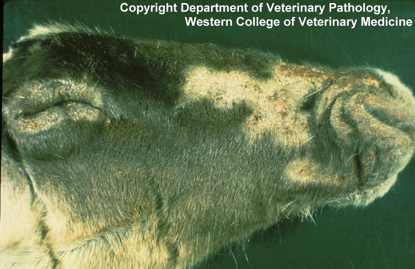
Hair loss and crusty skin
on the face of a caribou infected with Besnoitia. |
|
| Distribution |
|
Geographic: |
-
Several species of Besnoitia are found around the world that can
infect both wild and domestic herbivores. B. tarandi
is known to occur in BC.
|
|
Seasonality: |
-
Warmer months of the year; eggs passed in the feces of carnivores require
a warm
and moist environment to become infective.
|
|
| Hosts, Transmission and Life
Cycle |
| Hosts: |
-
Requires both an
intermediate host (in BC Besnoitia has only been
seen in caribou, but elsewhere has been reported in mule
deer) and
definitive host (carnivore: species in BC not known).
|
|
Transmission and Life Cycle: |
-
An
intermediate host ingests eggs that mature within the
gastrointestinal tract.
-
Within host cells, generations of asexual reproduction occur beginning
first in the walls of blood vessels followed by reproduction
in various organs and tissues forming relatively large
cysts (up to 1 mm in diameter).
-
When
cyst-containing tissues are
ingested by a
definitive carnivore host,
cysts break open within the
intestine and eventually differentiate into female and male
components and invade the tissues of the intestinal wall.
-
Eggs are produced and are
excreted in the feces and become infective once in the
external environment. Contaminated vegetation is ingested by
the herbivorous intermediate host and the cycle repeats.
-
Transmission between
intermediate hosts, independent of
definitive hosts, may occur through biting insects.
|
|
| Signs and Symptoms |
-
Clinical signs are observed only in
intermediate hosts and never in
definitive hosts.
-
Infected animals usually appear healthy and signs can vary.
Cysts observed in eyes of caribou
may not be present several days later.
-
A high density of
cysts on the skin can increase
the thickness of the skin while decreasing its elasticity, resulting
in the formation of cracks, allowing for
bacteria to enter and cause
infection.
-
Severe lesions have been
seen in captive animals with localized hair loss, fluid seepage and
hemorrhage, especially when large
numbers of
cysts are observed on joints of
the lower limbs, face and nasal cavity, and less often in the eye.
Skin,
subcutaneous tissue and the white of the
eye may look like sandpaper.
-
Thickening of skin within
the nasal passages can obstruct breathing.
-
Blockage of blood vessels
may be observed in B. tarandi infections.
-
The severe signs described
above have not been observed in wild
ungulates in BC.
-
When skinning the lower legs
of an infected animal, the
cysts can be observed as small
clear to white spheres.
Cysts are hard and have a slight
roughness that gives the underlying
connective tissue the appearance of being sprinkled with corn meal.
|
| Meat Edible? |
-
Meat is edible, cook well. DO NOT FEED
INFECTED MEAT TO DOGS.
|
| Human Health Concerns and
Risk Reduction |
-
There are no known human health concerns with Besnoitia infection.
|
| Samples for Diagnosis |
-
Submission of lower limb can be used for diagnosis, or examination of the
white of the eye.
|
| Similar Diseases |
|
|
| Further Reading |
-
Alaska Department of Fish and Game
– Besnoitiosis
-
Elkin B., Zamke R.L. 2001.
Common Wildlife Diseases and
Parasites in Alaska. Alaska Department of Fish and
Game. Anchorage, AK.
-
Leighton F.A., Gajadhar
A.A.. 2001. Besnoitia spp. and Besnoitiosis. Pp. 468-478 in
E.S. Williams, I.K. Barker (eds.),
Infectious Diseases of Wild
Mammals. 3rd Ed. Iowa State University Press. Ames, IA.
|
|
|