|
Baylisascaris (Raccoon Roundworm) |
 |
 |
 |
 |
|
| |
| Causative Agent |
-
Parasitic and
zoonotic disease of mammals and
birds caused by infection with the
roundworm (Nematode), Baylisascaris procyonis.
-
Immature (larval stages) of the worm migrate
through tissues and may cause extensive damage in susceptible hosts.
This is a trait shared by other
roundworms.
|
| Images |
|
Click on image to enlarge. |
 |
|
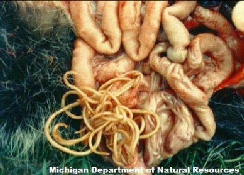
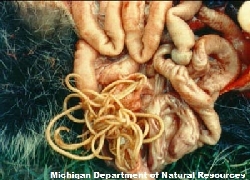
Raccoon roundworms are found
in the small intestines of infected animals. |
|
| Distribution |
|
Geographic: |
-
The distribution of B. procyonis mirrors that of raccoons (Procyon
lotor).
-
Raccoons are regularly found in the Lower
Mainland, southern BC and Vancouver Island, although their
range is expanding.
-
A recent study in southwestern BC indicated that the number of raccoons
infected with B. procyonis was 61%.
-
As raccoons are increasingly being brought as pets to new locations, the
geographic range of B. procyonis will continue to
expand.
|
|
Seasonality: |
-
New infections begin in young raccoons that have ingested infective eggs
of B. procyonis in late spring and early summer.
|
|
| Hosts and Life
Cycle |
-
Two alternate life cycles occur: one in raccoons, and the
other in susceptible, incidental (abnormal) hosts.
-
For a visual description of life cycles described below,
please visit the
US Centre for Disease Control and Prevention.
-
Definitive Host:
-
Adult worms are found in the small intestine of the raccoon.
-
Disease resulting from migrating larvae is rarely observed in raccoons.
Although, when disease is detected, it is seen in young
raccoons more often than adults.
-
Adult worms produce eggs that are shed in the feces. This can amount to
millions of eggs released per day/raccoon.
-
Within a month in the external environment, larvae develop within the
eggs, which are then infective.
-
Eggs may persist in the environment for years and are resistant to common
disinfectants. Burning is said to be the most effective
method of destroying the eggs.
-
Infective eggs are ingested by susceptible young raccoons OR infection
may occur after eating another animal that has larvae in
its tissues. Larvae migrate via the bloodstream through
the liver to the lungs.
-
The larvae are then coughed up, swallowed, and mature into adults in the
small intestine.
- Abnormal Host:
-
Many mammals and birds have been reported as abnormal hosts, including
humans, woodchucks, red and grey squirrels, porcupines,
cottontail rabbits, and a number of species of
ground-foraging birds.
-
Ingestion of larvae or eggs results in infection.
-
Raccoons use communal sites for defecation; other animals that forage in
these areas, as well as humans coming into contact with
such sites, are potentially at risk for Baylisascaris
infection.
-
Larvae hatched in the gut of abnormal hosts may migrate erratically
through tissues, such as lung, liver, heart and, most
notably, in the eyes and central nervous system.
-
Larvae
encyst in muscle, liver or the
lungs.
-
Larval migration through the
brain of susceptible hosts causes extensive tissue
damage, resulting in severe neurological signs that
include imbalance, circling and abnormal behavior.
-
Central nervous system
damage has been reported in humans and a large number of
wild and domestic mammals and birds.
|
|
| Signs and Symptoms |
-
Like other
roundworms, B. procyonis are cylindrical and taper at both ends. Adult worms
are tan-white in color, measure 9-22 cm in length and 1 cm in
thickness.
- In raccoons:
-
Larval migration may cause localized areas of
inflammation and tissue damage or cause
damage due to blockage of the small intestine by adult worms.
B. procyonis infection otherwise seems to have no
detrimental effects on raccoons.
- In abnormal hosts:
-
There are usually no symptoms if the larval parasite does not enter the
brain.
-
Effects are usually correlated with the number eggs ingested, the number
of larvae entering the brain, extent of migration within the
brain, and size of the brain relative to the size of the larval
parasite.
-
Larvae may become
encapsulated in tissues; these
cysts are usually visible as
light-colored spheres, which are 1-2 mm in diameter.
-
Clinical signs in small mammals include:
-
depression;
-
lethargy;
-
nervousness;
-
rough coat;
-
tremors in the front paws;
-
head or body tilts: slight
at first, progressing to worse;
-
falling over;
-
circling;
-
posterior
paralysis;
-
blindness;
-
laying on its
side.
-
Clinical signs in birds include:
-
poor grip reflexes;
-
incoordination;
-
inability to fly or loss of
flight control;
-
falling;
-
wing and leg
paralysis.
-
Clinical signs in humans include:
-
skin irritation from larval
migration within the skin;
-
eye and brain tissue damage
due to the random migration of larvae;
-
individuals may experience
nausea, a lethargic feeling, incoordination and loss of
eyesight.
|
| Meat Edible? |
-
Raccoon meat is generally not consumed by humans. If a raccoon is to be
skinned, proper protective gear should be worn (gloves, coveralls)
and good hygiene should be practiced.
|
| Human Health Concerns and
Risk Reduction |
-
Baylisascaris infection in humans may
cause severe damage in the eyes and brain, and in extreme cases,
death.
-
Minimizing the potential
exposure of people to raccoon feces is the best risk reduction
measure.
-
Exclusion of raccoons from
areas of human habitation is warranted, as is careful attention to
hygiene, particularly of children, in high-risk areas.
-
Wildlife rehabilitators,
animal shelter workers and others who may come in contact with
raccoon feces on a regular basis need to take particular care in the
handling and disposal of raccoon feces. Additionally, these
organizations should deworm all raccoons that come under their care,
although this is not guaranteed to remove all parasites.
|
| Samples for Diagnosis |
-
Infection with Baylisascaris may be confirmed by finding eggs in
the fecal material of live raccoons.
-
Roundworms found in the intestines of
raccoons should be submitted to determine if they are B.
procyonis.
-
Tissues of hosts other than
raccoons that contain small
cysts should be submitted to determine if B.
procyonis larvae are present.
|
| Similar Diseases |
-
Neurological symptoms
are very similar to
rabies and other wildlife diseases that affect the central
nervous system.
-
Other disease agents which may elicit similar
neurological symptoms include:
|
| Further Reading |
-
BC Centre for Disease Control – Raccoon
Roundworm
-
Canadian
Medical Association Journal – Raccoon
Roundworm
-
Canadian Cooperative Health Centre –
Raccoon Roundworm (PDF file)
-
Michigan Department of Natural Resources –
Raccoon Roundworm
-
Canadian Cooperative Wildlife Heath Centre. 1995. Baylisascaris
procyonis Larval migrans. Pp. 45-47.
Health Risks to Wildlife
personnel: Hazards from Disease-causing Agents. Canadian
Cooperative Wildlife Heath Centre, Western College of Veterinary
Medicine, University of Saskatchewan. Saskatoon, SK.
-
Ching H.L., Leighton B.J., Stephen C. 2000. Intestinal parasites of
raccoons (Procyon lotor) from southwest British Columbia.
Canadian Journal of Veterinary
Research 64: 107-111.
-
Coates J.W., Siegert J., Bowes V.A., Steer D.G. 1995. Encephalitic
nematodiasis in a Douglas squirrel and a rock dove ascribed to
Baylisascaris procyonis.
Canadian Veterinary Journal 36: 566-569.
-
Kazacos K.R. 2001. Baylisascaris procyonis and related species.
Pp. 301-341 in W.M. Samuel, M.J. Pybus, A.A. Kocan (eds.),
Parasitic Diseases of Wild
Mammals. 3rd Ed. Iowa State University Press. Ames, IA.
|
|
|